[ad_1]
The following chest x-rays are from an 11 year old presenting with fever and a cough. What can you see?

click to enlarge
click to enlarge
[peekaboo_link name=”Answer”]Answer[/peekaboo_link]
[peekaboo_content name=”Answer”]
The PA view shows the silhouette sign – the medial half of the left dome of the diaphragm is obscured by the consolidation process. There is left lower lobe pneumonia.

click to enlarge
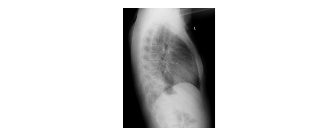
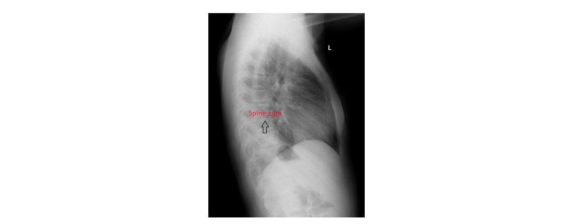
On the lateral view, the spine sign is positive. There is a triangular opacity over the lower vertebral bodies. Normally, the lower thoracic vertebral bodies appear darker compared with the vertebrae in the mid and upper thorax. This is because of the increased soft tissue densities over the upper and mid thoracic vertebra.
click to enlarge
This child has pneumonia and should be treated with antibiotics.
[/peekaboo_content]
[ad_2]
Source link