[ad_1]
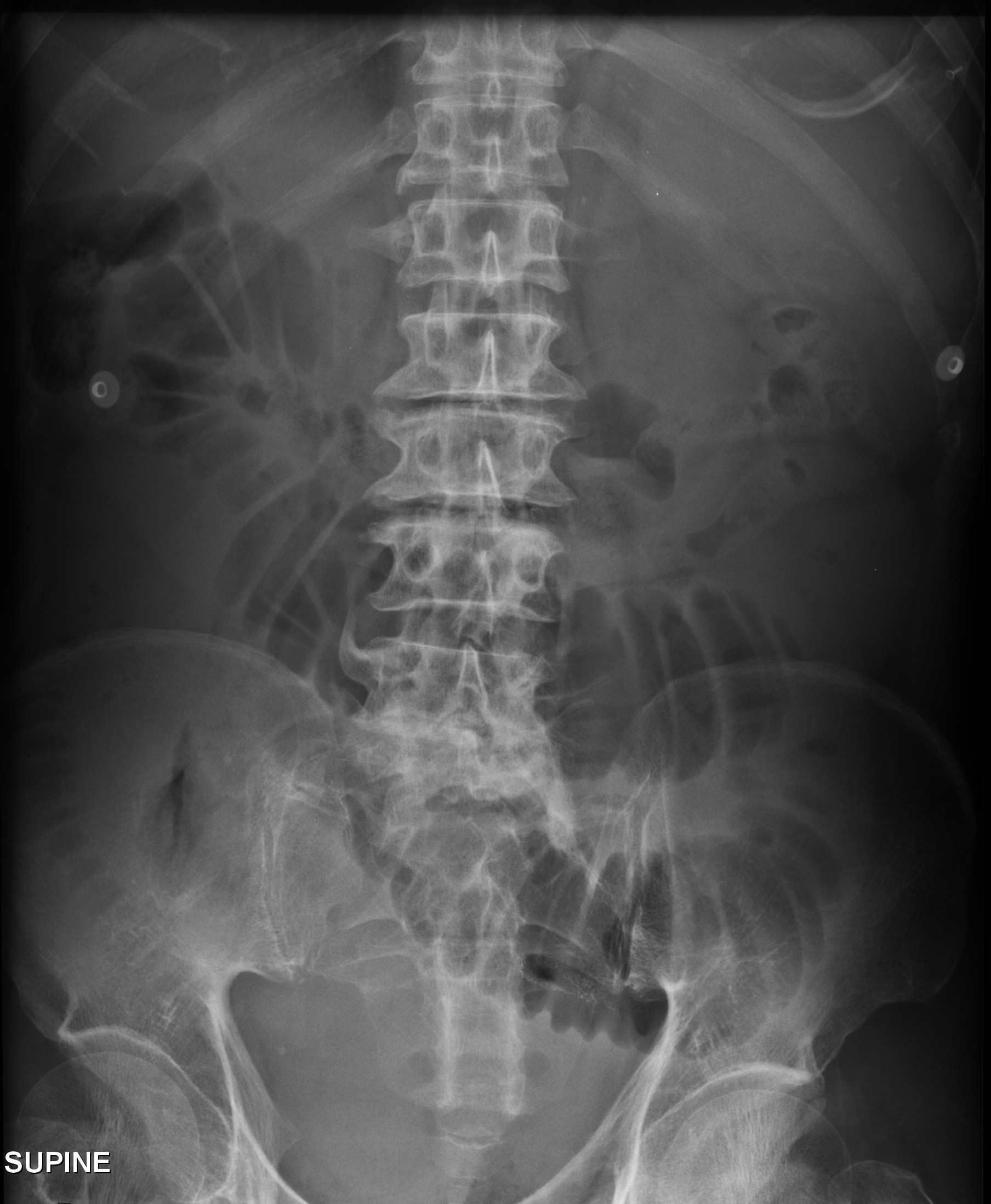
This supine abdominal x-ray is from a 60 year old patient who has presented to the ED with abdominal pain and vomiting. Anatomically, which part of the small bowel is obstructed?
click to enlarge
[peekaboo_link name=”Answer”]Answer[/peekaboo_link]
[peekaboo_content name=”Answer”]
The supine abdominal x-ray shows likely distal jejunal obstruction. The dilated jejunal loops are centrally located and have a ‘stacked coins’ appearance to them and this is typical of jejunal loop dilatation. The diameter of the dilated loops is up to 4.0 cm.
The vavulae conniventes cross the entire width of the jejunum and are responsible for the stack of coins appearance. Distended ileal loops are featureless. Colonic distension is peripheral in location and has haustra which do not cross the entire width of the bowel.
[/peekaboo_content]
[ad_2]
Source link