[ad_1]
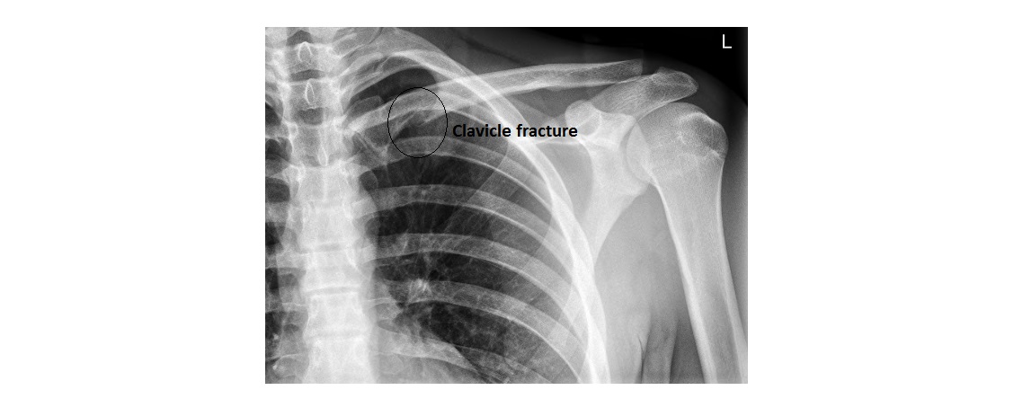
This is the shoulder x-ray of a 16 year old patient who has sustained injury following a fall onto his left shoulder while playing rugby. Patient is not tender over the head or neck of humerus. What can you see?

click to enlarge
[peekaboo_link name=”Answer”]Answer[/peekaboo_link]
[peekaboo_content name=”Answer”]
The shoulder joint area appears normal and the lateral view was fine too.
The x-ray shows fracture in the medial third of clavicle. The superior cortex is intact but the inferior cortex is broken.
click to enlarge
For some reason this patient did not have a proper AP view of his clavicle to begin with. Although most clavicle fractures are easy to spot, some may not be visible on the standard AP view. If clinical index of suspicion is high, then additional 45 degree cephalic tilt view has to be requested.
[/peekaboo_content]
[ad_2]
Source link