[ad_1]
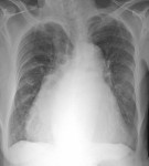
The following erect PA chest x-ray is from a 65 year old female patient with exertional dyspnoea and orthopnoea. There is an obvious cardiomegaly. What is the underlying valvular pathology?

click to enlarge
[peekaboo_link name=”Answer”]Answer[/peekaboo_link]
[peekaboo_content name=”Answer”]
This patient had mixed mitral valve disease diagnosed on echocardiogram.
The 3 abnormalities suggestive of left atrial enlargement on the chest xray are:
- Double right heart border
- Splaying of the carina
- Straightening of the left heart border due to enlargement of the left atrial appendage.

click to enlarge

click to enlarge
The double right heart border – formed by the edge of the enlarged left atrium. An enlarging left atrium bulges to the right. On the rigth side, borders of the right and left atria can be distinguished as the left atrial border passes medially towards the spine before fading, whereas the right atrial border is limited below by entry of IVC.
Splaying of the carina – the enlarging left atrium elevates the left main brochus and causes widening of the carinal angle (should not be more than 75 degrees).
Both of the above findings are late signs of left atrial enlargement whereas straightening of the left heart border occurs earlier.
References:
- Grainger and Allison’s diagnostic radiology
- Mettler: Essentials of radiology
[/peekaboo_content]
[ad_2]
Source link