[ad_1]
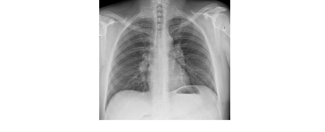
This is the chest x-ray from a 40 year old man who has presented to the emergency department with a cough and mild dyspnoea. Anything obvious on the x-ray?
Click to enlarge
[peekaboo_link name=”Answer”]Answer[/peekaboo_link]
[peekaboo_content name=”Answer”]
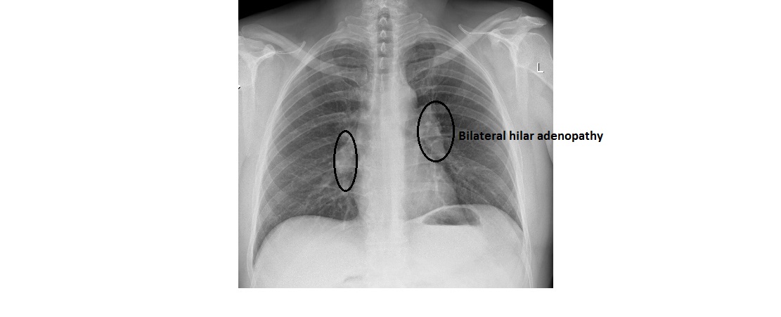
The chest x-ray shows clear lung fields.There is no pleural effusion and no pneumothorax. The left diaphragm is higher compared to the right (long standing finding).
The notable feature on this xray is lobulated bilateral hilar enlargement. There is symmetrical (more or less) bilateral hilar adenopathy. This patient was later diagnosed with sarcoidosis.
click to enlarge
Based on chest x-ray findings, pulmonary sarcoidosis is classified into 4 stages:
- Bilateral hilar adenopathy but otherwise clear lungs.
- Bilateral hilar adenopathy and diffuse reticulonodular disease pattern in the lungs.
- No lymphadenopathy but diffuse coarse reticulonodular pattern.
- Scarring/fibrosis in the lung parenchyma.
[/peekaboo_content]
[ad_2]
Source link