[ad_1]
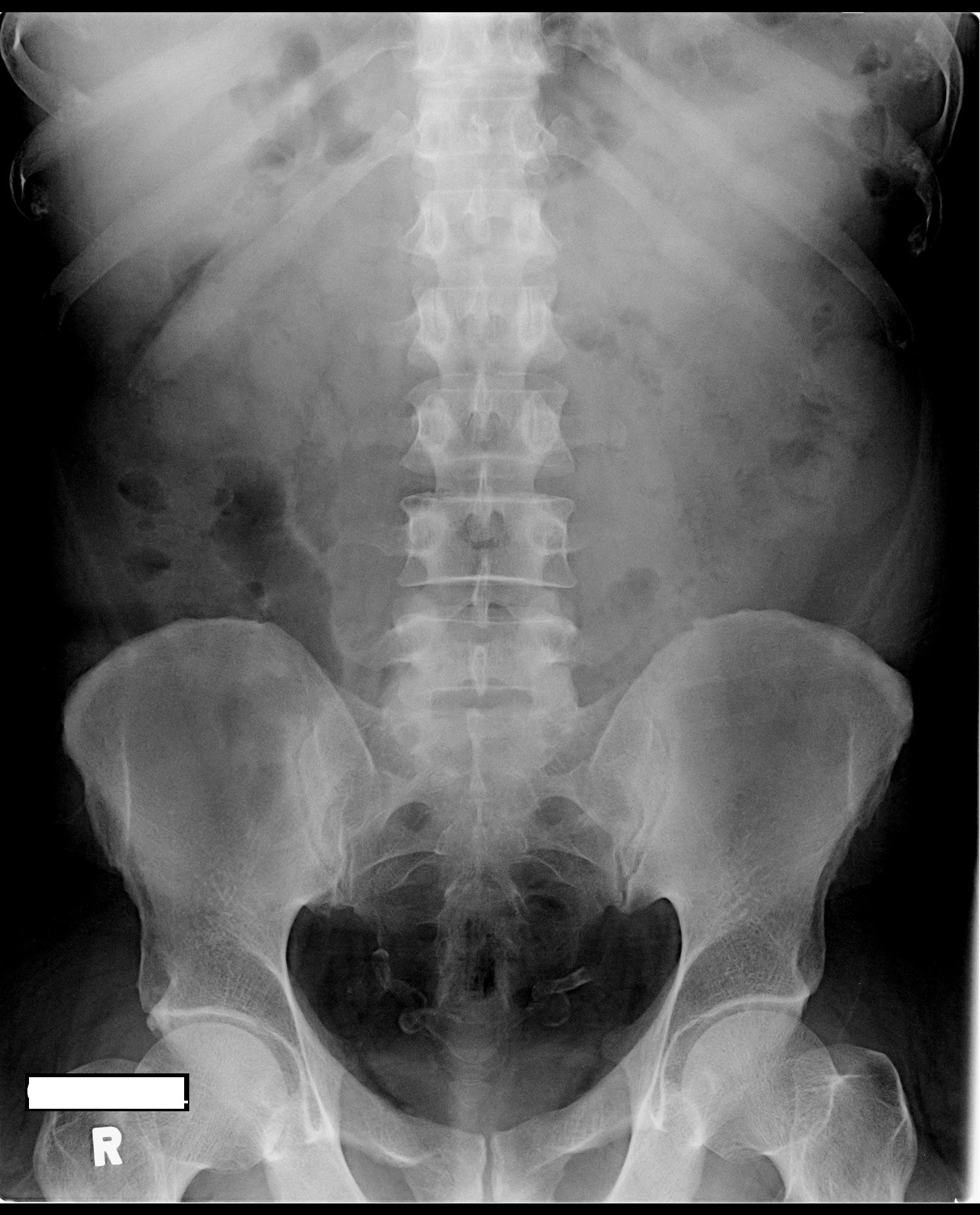
This KUB xray is from a 55 year old diabetic man who presented to the ED at about 3am with unilateral colicky abdominal pain.You suspect he is suffering from ureteric colic. What does the xray show?
[peekaboo_link name=”Answer”]Answer[/peekaboo_link]
[peekaboo_content name=”Answer”]
The x-ray does not show any radio-opaque calculus in the kidney ureter bladder area. There is an incidental finding of bilateral calcified tubular serpentine structures in the pelvic area; this represents calcified vas deferens (nothing related to emergency really).
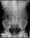
Causes are long standing diabetes as is the case with this patient. Other causes can include sexually transmitted diseases such as syphilis, gonorrhoea or local chronic infections such as tuberculosis and schistosomiasis.
[/peekaboo_content]
[ad_2]
Source link