[ad_1]
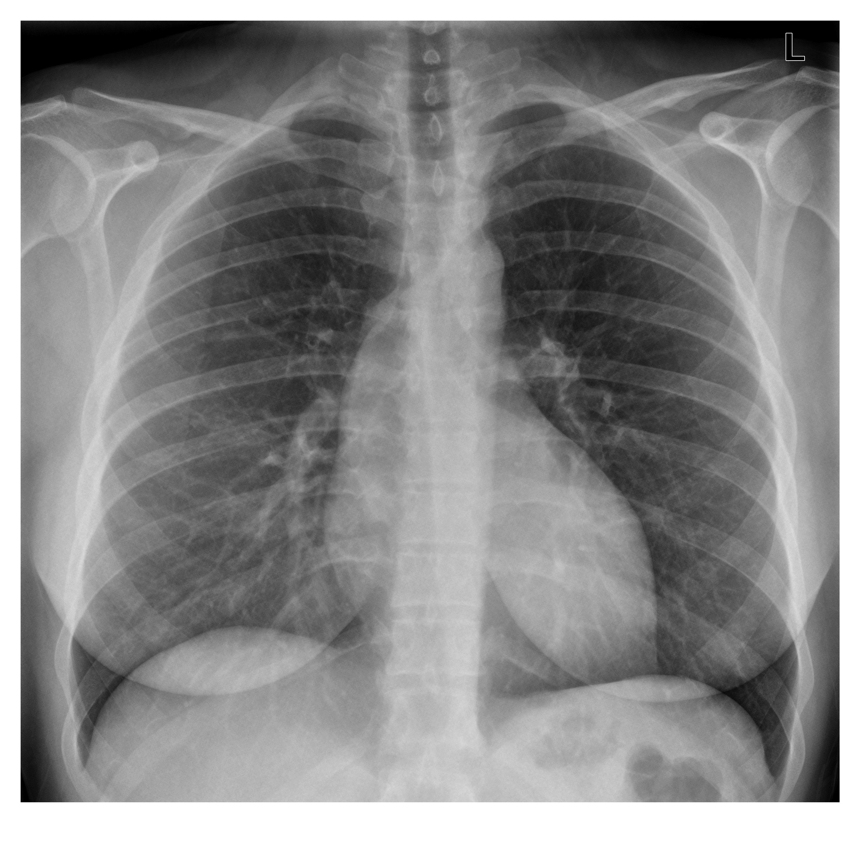
A 34 year old woman who is 3 days post-partum has presented with a sudden onset of central chest pain at 3 am. She has no previous medical illness. As part of investigations, a chest x-ray has been done and is as follows. What is seen on the x-ray? (It is very subtle)
Click to enlarge
[peekaboo_link name=”Answer”]Answer[/peekaboo_link]
[peekaboo_content name=”Answer”]
At first glance, the chest x-ray appears normal. The lung fields are clear; there is no pneumothorax to account for the sudden onset of pain. However, there is a subtle finding on the cardiac outline which raises the possibility of proximal aortic dissection.
There is a diffuse bulge along the superior aspect of the right heart border, causing focal widening of the mediastinum. This finding is due to aortic root dilatation associated with dissection (Stanford A).

Click to enlarge
This patient had a confirmed proximal aortic dissection on CTA. A slice of axial post contrast CT chest is as follows and shows intimal flap in the mildly dilated ascending aorta (diameter is 4.5 cm).

Click to enlarge
[/peekaboo_content]
[ad_2]
Source link