[ad_1]
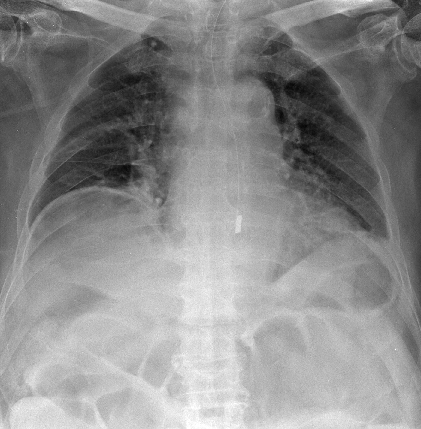
This is the supine chest/abdominal x-ray of an elderly patient who has presented with abdominal distension with pain and vomiting. Apart from the very obvious (malpositioned nasogastric tube), what interesting radiological sign can you see?
Click to enlarge
[peekaboo_link name=”Answer”]Answer[/peekaboo_link]
[peekaboo_content name=”Answer”]
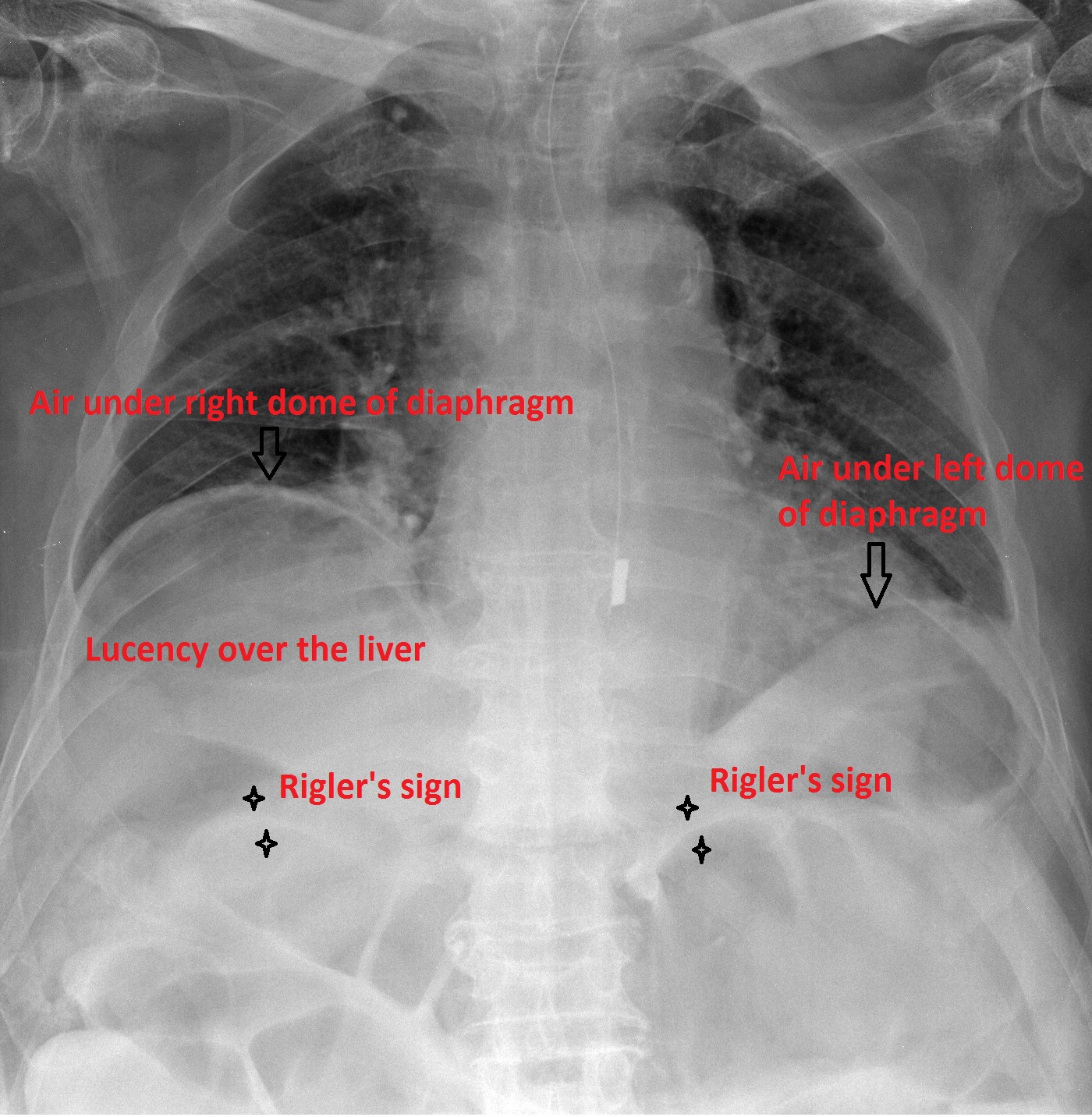
X-ray shows a large amount of free intraperitoneal air under both domes of diaphragm and also lucency over the liver. Normally, the hepatic shadow on a x-ray should not have any overlying luceny. The radiological sign seen is the Rigler’s sign which is also called the double wall sign.
Rigler’s sign is present when the bowel wall is clearly visible as a white line because of gas within the bowel as well as outside the bowel wall.
Click to enlarge
[/peekaboo_content]
[ad_2]
Source link