[ad_1]
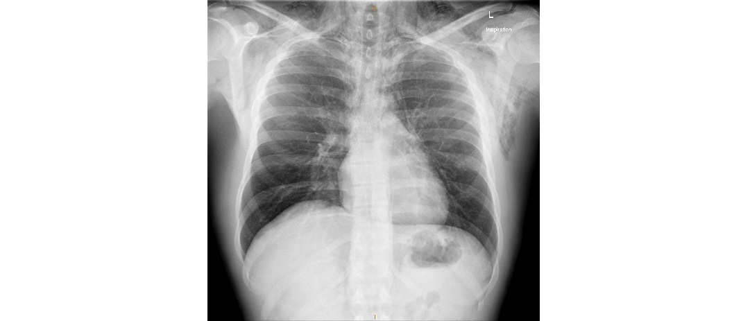
You have just seen a 20 year old patient who presents with dyspnoea and chest pain. He has been partying hard on New Year’s Eve and admits to ‘snorting’ something while at the party. The chest x-ray is as follows; what interesting radiological sign can you see?
Click to enlarge
[peekaboo_link name=”Answer”]Answer[/peekaboo_link]
[peekaboo_content name=”Answer”]
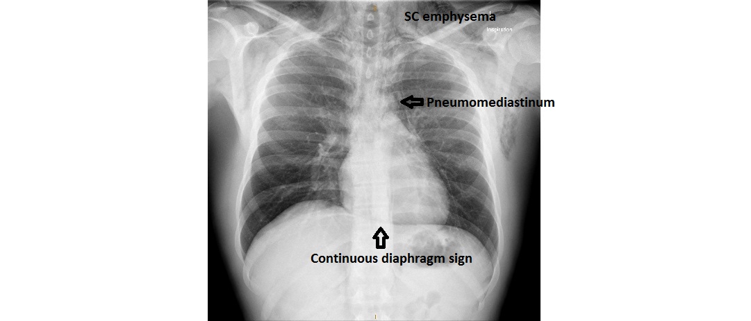
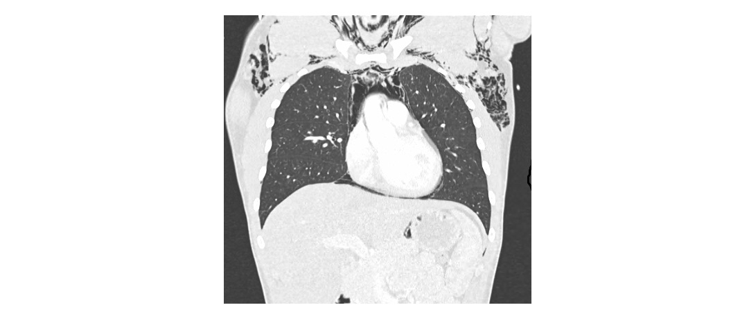
There is an obvious pneumomediastinum with extensive subcutaneous emphysema visible on the x-ray. The interesting radiological sign is the continuous diaphragm sign which is due to the stripe of air interposed between the diaphragm and the pericardium. Normally the central diaphragm is obscured by the heart.
Click to enlarge
Click to enlarge
Continuous diaphragm sign can be due to pneumomediastinum (lucent stripe above the diaphragm) or pneumoperitoneum (lucent stripe below the diaphragm).
It can be the only sign of pneumomediastinum especially in supine trauma patients.
Thanks to Dr. Yuresh Naidoo for the images.
[/peekaboo_content]
[ad_2]
Source link