[ad_1]
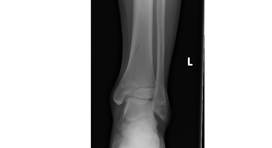
These ankle x-rays are from a 15 year old patient who has injured his left ankle while skateboarding and is unable to weight bear since injury. What can you see?
Click to enlarge
[peekaboo_link name=”Answer”]Answer[/peekaboo_link]
[peekaboo_content name=”Answer”]
The AP and the mortise view x-rays show what looks like a Salter and Harris type III fracture of distal tibial growth plate with fracture line involving the medial aspect of the distal tibial epiphysis with intra-articular extension.
On the lateral ankle view, the fracture line runs through the distal tibial diaphysis, metaphysis and epiphysis indicating a type IV injury. There is widening of the growth plate of the anterior tibia on the lateral view. There is also significant ankle joint effusion.
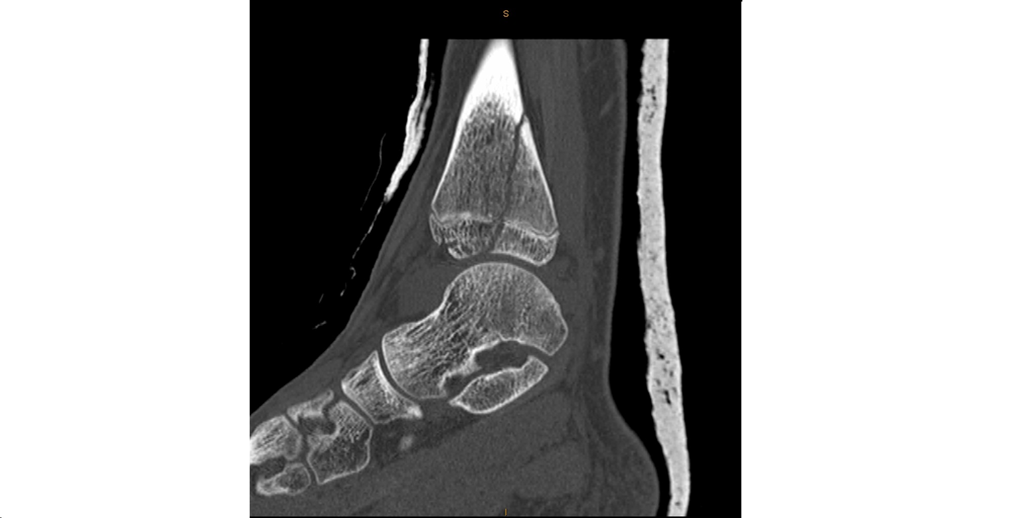
CT scan of the ankle confirmed Salter-Harris type IV fracture.
Click to enlarge
There is a mnemonic SALTR for the 5 types of fractures involving the growth plate in children and details can be found on the internet.
[/peekaboo_content]
[ad_2]
Source link