August 25, 2025
3 min read
Key takeaways:
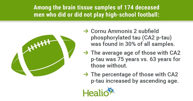
- CA2 p-tau was found in 30% of all samples.
- The average age of those with CA2 p-tau was significantly older vs. those without.
- The percentage of samples with CA2 p-tau increased by ascending age groups.
No significant associations were found between tau levels found in a specific area of the hippocampal region of the brain and participation in high-school football, according to research published in the Journal of Alzheimer’s Disease.
Hyperphosphorylated tau (p-tau) accumulates in the Cornu Ammonis 2 subfield (CA2) of the hippocampus as individuals age, in association with the early onset of Alzheimer’s disease, Rudolph J. Castellani, MD, professor of neuropathology at Feinberg School of Medicine, Northwestern University, and colleagues wrote.

“The [Boston University]/Concussion Legacy Foundation (BU/CLF) brain bank studies specifically recruit only deceased athletes and only deceased athletes who had mental health or neurological problems during life,” Castellani told Healio.
Castellani and his colleagues examined cases for CA2 tau presence regardless of repetitive head impact or sport history, which he said can more accurately assess risk and causal inferences.
As such, the researchers conducted a case control study to determine potential associations between p-tau in the hippocampal CA2 region and history of participation in high-school football.
“Tau in CA2 was noted as a ‘supportive’ feature of [chronic traumatic encephalopathy] in a 2016 consensus article,” Castellani said regarding the focus on this specific region of the brain. “It was later reported that CA2 tau was a benign aging-related change. The neuropathology community seems to be on both sides of this issue.”
Working under the hypothesis that there would be no statistically significant difference in CA2 p-tau between participation groups, Castellani and colleagues analyzed brain tissue samples from 174 deceased men (median age at death, 65 years) obtained from the Lieber Institute for Brain Development, a nonprofit brain research center in Baltimore. Of these, 126 belonged to those with no known history of participation in contact sports and 48 who participated in high-school level football.
“Lieber Institute does not specifically recruit athletes, so it does not have the selection bias inherent in BU/CLF studies,” Castellani said.
The samples were additionally subdivided by age into 10-year blocks (50 to 59 years, 60 to 69 years, 70 to 79 years, 80 years and older) for both groups.
Preferentially CA2 p-tau immunolabeling was positively identified if seen at a low magnification and included the presence of neuronal cell pretangles, both with or without neurofibrillary tangles, according to the researchers.
P-tau and amyloid-beta were also quantified using modifications of the standard Braak staging system as well as the Consortium to Establish a Registry for Alzheimer’s Disease (CERAD).
No significant associations were found between those who played football and preferential CA2 p-tau, although its presence was detected in 29.9% of the samples overall. By group, 27.8% of those who played high-school football and 35.4% of those who did not had preferential CA2 p-tau.
Data further showed an older average age for those with vs. those without preferential CA2 p-tau (75 years vs. 63 years), with the percentages of those having CA2 p-tau increasing by ascending age (50 to 59 years, 11.1%; 60 to 69 years, 13.6%; 70 to 79 years, 54.1%; 80 years and older, 75%).
In both univariate and multivariate logistic regressions, preferential CA2 p-tau was significantly more likely to appear in individuals who were older (OR=3.42 and 3.23, respectively) and in those with greater modified CERAD scores (OR=1.78 and 1.48).
The researchers additionally reported that approximately half of the samples were rated modified Braak stage I (47.1%) and modified CERAD stage 0 (52%), indicating minimal or no involvement with the hippocampus.
“This is typical of CTE neuropathology in the 21st century: subtle and nuanced microscopic findings in the postmortem brain that have no relevance or significance to living human beings,” Castellani told Healio.
References:
For more information:
Rudolph J. Castellani, MD, can be reached at: neurology@healio.com.


