By Trina Augustin MD
In partnership with…
- Gabriel Ortiz Jaimes, MD (Interventional Pulmonology Fellow- Mayo Clinic, Rochester, MN)
- John Mullon, MD (Interventional Pulmonologist – Mayo Clinic, Rochester, MN)
- Ryan Kern, MD (Interventional Pulmonologist – Mayo Clinic, Rochester, MN)
Peer reviewed by:
- Lewis McLean, Intensivist with special interest/training in cardiovascular critical care and MCS (NSW, Australia)
- Deepa Patel, Intensivist and Emergency Medicine Physician with special interest in Ventilator mechanics (Emory University, Atlanta, GA)
Podcast Guests
- Trina Augustin – CV Intensivist and ECMO maven. Editor, CV-EMCrit
- Gabriel Ortiz Jaimes, MD – Trained in Internal Medicine (Universidad Nacional de Colombia and University of Texas Health Sciences Center at San Antonio). Pulmonary, Critical Care Medicine, and Interventional Pulmonology at Mayo Clinic in Rochester Minnesota. Interest in benign and malignant airway and pleural disease.
Why are we diving into persistent air leaks (PAL) with bronchopleural and alveolopleural fistulas on CV EMCrit?
I kept getting VV ECMO consults on them and found myself managing these complex cases alongside my interventional pulmonary colleagues. Naturally, I took the opportunity to nerd out—mixing transpulmonary pressures and mechanical circulatory support—while wrestling with the nuanced challenge of gas exchange in the sickest subset of these patients.
But first—I teamed up with my phenomenal, world-class interventional pulmonary colleagues, who generously shared their approach to managing persistent air leaks in Parts I & II. Part III dives into the sickest subset: patients with PAL who require mechanical ventilation and VV ECMO to maintain gas exchange and facilitate fistula healing.
Part I:
Persistent Air Leak (PAL): Definitions, Severity, and First-Line Management
We toss around “persistent air leak” (PAL) like it’s universally defined — it’s not. The literature is fragmented, and definitions vary widely, especially across surgical vs. medical domains. This complicates incidence stats, outcome tracking, and management thresholds.
📚 Want deep dives? Start with these reviews: Mayo’s take (PMID: 29605207) + another excellent review (PMID: 28267436).
👁️🗨️ Key Terms
- Alveolo-pleural fistula (APF): Communication from lung parenchyma (distal to subsegmental airways) to pleural space
- Bronchopleural fistula (BPF): Communication from airways proximal to the subsegmental level (mainstem, lobar, segmental) to pleural space — potentially visible on standard bronchoscopy.
- Persistent Air Leak (PAL): The clinical expression of APF or BPF—caused by continued air flow from the endobronchial tree to the pleural space. Evidenced by continued bubbling in the water seal chamber or recurrent pneumothorax despite chest drainage.
PAL = air leak that sticks around. But for how long? That depends.
- Classic threshold: more commonly >5 days (range 3-7 days) after chest tube insertion (initial leaks are common post-lung resection).
- Society guidance (pre-dating pleurodesis, endobronchial valve era):
- ACCP/BTS: Consider PAL if leak persists >48hrs after pleural drainage (PMID: 11171742).
- Modern guidelines (ERS/EACTS 2024, BTS 2023): Still largely unchanged (PMIDs: 38806203, 37433578)
🧠 Bottom line: The longer the leak, the higher the risk — especially post-op. PAL has been linked with up to 30% mortality in surgical series.
Basic principle: The timing of the air leak within the respiratory cycle reflects its severity and the pleural pressure needed to sustain flow across the defect
🫁 Cerfolio Classification (PMID: 9875779)
- Grade 1: Bubbling only on forced exhalation (e.g. cough)
- Grade 2: Bubbling during normal expiration
- Grade 3: Bubbling during inspiration
- Grade 4: Continuous bubbling (insp + exp)
- 💡 Most leaks are Grade 1–2. Grades 3–4 = larger, more proximal defects (think BPF, PPV patients).
📊 Leak Volume ≠ Guesswork Anymore
- Column-based systems (e.g. Atrium): Count the columns = semi-quantitative leak assessment. The number of “columns” of leak present can be followed longitudinally to determine progression or resolution of the leak
- Digital drainage systems (e.g. Thopaz Medela): Quantitate airleak flow.
- Spontaneous pneumothorax (SP) leaks >100 mL/min = higher failure of conservative therapy (PMID: 30355640)
🔬 Etiology: Why Leaks Linger
🫁 Any etiology causing a pneumothorax can lead to PAL, but higher risk with…
- Abnormal underlying parenchyma → prone to APF (more common in lung disease)
- Disrupted proximal airways → risk of BPF (more common post-surgery/airway instrumentation or in malignancy) (In press DOI: 10.1016/j.ccm.2025.02.015 )
- Instrumentation, malignancy, barotrauma → high risk of conversion to PAL
✂️ The Big Buckets: Surgical vs. Spontaneous + “Other Etiologies”
1) Post-Surgical PAL
- Highest rates after lung volume reduction surgery (LVRS):
- 46% in clinical trials
- Up to 90% in some series (severely diseased emphysematous lungs)
- Compare that to:
- Lobectomy: 8–10%
- Segment/wedge resection: 3–8%
🧠 Risk factors: Emphysema, poor PFTs, smoking, steroid use, diabetes, pleural adhesions, upper lobe resections (PMIDs: 21871301, 16798215). Significantly increases mortality (PMID 19853117)
2) Spontaneous Pneumothorax (SP)
Divided by baseline lung:
- Primary SP: Healthy lungs (usually young males, subpleural blebs)
- Secondary SP: Diseased lungs or smokers>age 50 — much higher risk of PAL
- Think COPD, ILD, cystic lung diseases, infections
- One series: 34.6% incidence of PAL in secondary SP (PMID: 9713636)
3) Other Etiologies: Trauma & Iatrogenic PAL
- Includes chest tube placement, biopsies, mechanical ventilation, and barotrauma.
- Barotrauma is a common cause in ICU settings
⚖️ Management: Drain, Wait, Watch (Then Escalate)
🧰 Chest Tubes 101
- Surgical PAL: typically, will have large-bore tubes
- Medical PAL: Small-bore (“pigtails”) preferred — lower complication rates, shorter drainage duration (PMID: 29452099)
- ⚠️ Exception: Patients on positive pressure ventilation, high-volume leaks, barotrauma, large proximal BPF — may need large-bore or multiple tubes as they may develop tension physiology with smaller drains
- Monitor for progressive SQ emphysema which can clue you in to an uncontrolled air leak even if PTX is not expanding
⏳ Just Drain & Observe: Not Passive, Just Smart
- Secondary SP PAL: 61% resolve in 7 days, 79% by day 14 (PMID: 9713636)
- Primary SP: Even higher resolution rates
- Takeaway: Waiting works. If stable — don’t rush.
🔌 Apply Suction: Yes or No, the controversies continue.
- Post-surgical data: Early transition (day 2) to water seal is significantly superior to suction in terms of resolution of the air leak (PMID 11383809)
- SP & iatrogenic leaks: No time-to-resolution benefit in water seal vs suction (PMIDs: 17189116, 7071793)
- Current Guidelines (ERS/BTS): No strong evidence for routine suction
- Our (authors) opinion: An early transition to water seal is likely safe and could have favorable aspects
🚶 Outpatient Strategy: Heimlich Valves
If they tolerate water seal, they can often go home.
- Safe + effective: >60% have tube removed <1 month and average chest tube durations <10 days (PMID 8607667)
- Low complication rates (empyema the main concern) (PMID 19463579)
Next up in Part II: Pleurodesis, bronchoscopic tricks of the trade, and more
🔧 Pleurodesis: Forcing the Seal
Pleurodesis = induced permanent adhesion of the visceral and parietal pleura.
The goal? Eliminate the pleural space and stop the leak — permanently. It can be achieved surgically or medically, and is a cornerstone strategy for managing PALs.
1) Surgical Pleurodesis
- Mechanical abrasion or talc poudrage during operative intervention.
- Typically performed in planned or ongoing surgical management of PALs.
- Results in intense pleural inflammation → permanent adhesions.
2) Medical Pleurodesis
- Induced by the instillation of agents through the chest drain, capable of inducing pleural adhesion or inflammation
- Two most common approaches:
- Autologous blood patch pleurodesis (ABPP)
- Talc slurry pleurodesis (PMID: 39193457)
🧠 But first, Key Consideration Before Pleurodesis:
✅ Ideal: Patient tolerates clamping (no tension physiology, no desaturation)
🔄 Backup: Elevate drain to chest height while on water seal (“the loop”) to allow egress of air but retain the pleurodesing agent in the chest
❌ Poor candidates: Patients who can’t tolerate either
Ideal retention time varies (30–120 min), but longer = better adhesion.
Medical Pleurodesis continued…
1)🩸 ABPP: Seal with the Patient’s Own Blood
The theory: Blood coats the visceral pleura, forms a clot over the defect, and promotes localized inflammation + sealing.
🩸 How to do it:
- Fresh venous blood via peripheral stick
- Instill 1–3 mL/kg (Mayo practice), 100 mL shown to be superior to 50 ml, potentially earlier resolution (PMID: 17320580)
- Typically clamp or loop the chest tube on water seal for 30–120 min
📈 Efficacy:
- Success rates up to 80% (PMID: 3411599)
- Data is heterogeneous (PMID: 29672817, 22459543, 34477307), but consistently shows:
- ↓ air leak duration
- ↓ chest tube duration
- ↓ hospital stay
- 🔬 Works across scenarios: surgical or spontaneous PAL
- Can be combined with bronchoscopic therapies (e.g., valves) (PMID: 30904909)
Guidelines:
- ERS 2024: Consider in SSP patients unsuitable for surgery
- BTS 2023: No strong recommendation, but acknowledges shorter LOS with ABPP
Figure: The Mayo clinic blood patch protocol in surgical patients as below (PMID 34277031)
2) ⚗️ Talc Pleurodesis: 🔥 Talc is a potent pleural irritant:
- Commonly used in malignant pleural effusions, also can be used for PAL
- Doses: 4–12 g
- Rapid inflammation, adhesions within days
- IT CAN HURT: Requires aggressive analgesia pre & post
💉 Instilled as slurry (mixed talc and saline) through the chest tube
📊 Efficacy in PALs:
- Proven efficacy in surgical PALs — ↓ reintervention, earlier chest tube removal (PMID: 9875779)
- Also effective in SP-related PALs (PMID: 30863586)
🔪 Surgical Management: The Original Fix
- Before pleurodesis or advent of bronchoscopic techniques, surgery was the only avenue available. It still plays a major role — especially for post-op PALs, where it’s often first-line.
- Early BPF (<14 days) post pneumonectomy, goal is expedited surgical management to repair fistula (optimize/revise stump & prevent pleural spillage and infection
- Late BPF (>14 days) post pneumonectomy; primary BPF surgical closure less likely successful & more likely to have a mature BPF tract and possible emphysema. Focus is 1) drain pleural space & control infection, typically chest tube +/- decortication 2) obliterate pleural space
- Multiple surgical strategies and techniques are available but are beyond the scope of this review.
🧰 Big Picture: Surgery offers…
- Bronchial stump revision with coverage of proximal defects with Muscle/vital tissue flap coverage
- Completion resections (completion lobectomy, pneumonectomy) with surgical coverage techniques for extensive leaks
- Decortication and multiple techniques to try to control/drain pleural infection
🧠 Efficacy: (PMID 16798230, 29109057)
✅ >80% leak resolution
✅ <20% recurrence
🚨 But: Requires VATS or thoracotomy → significant morbidity, often not feasible in high-risk or frail patients.
🫁 Bronchoscopic Management: From Diagnosis to Fix
In recent years, bronchoscopic tools have moved from diagnostic adjuncts to frontline therapies. Techniques like air leak localization and the use of endobronchial one-way valves (EBVs) have transformed bronchoscopy into a core strategy for managing both medical and surgical PAL. In practice, it’s common to see a mix of bronchoscopic and non-bronchoscopic interventions used in combination to get leaks under control
1)🕵️♂️Bronchoscopic Leak Localization: Where’s the Hole?
- Direct bronchoscopic airway inspection
-
- Most effective at locating major stump and anastomotic dehiscence in surgical patients as well as proximal BPFs.
When the defect is not evident in the proximal airways on direct inspection, two additional bronchoscopic techniques can be invaluable to localize distal bronchial and parenchymal leaks to determine the location for interventions
- Balloon Occlusion Techniques
-
- Use a Fogarty catheter (most commonly) to sequentially block lobar/segmental bronchi (PMID 16354867, 29605207)
- Watch for leak reduction or resolution via the chest drain
- Complete cessation = target segment found
- 🧬 Note: some parenchymal air leaks (often associated with malignancy, infection, trauma) may have Collateral ventilation with other segments/lobes and often prevent complete air leak resolution → may need multi-segment occlusion
- Dye Instillation
- Instill diluted methylene blue via pleural drain
- Observe airways during bronchoscopy for dye egress → guides target location for future interventions
(PMID: 29605207, 28937444)
2) 🛑 Bronchoscopic Management: “The MVP”: Endobronchial Valves (EBVs)
A game changer in PAL management — especially non-surgical or high-risk patients.
Two unidirectional endobronchial devices FDA-approved systems for bronchoscopic “lung volume reduction in emphysema patients”:
- Spiration (Olympus) – umbrella EBV system, sizes 5–9 mm diameter, lengths 8-12 mm
- Zephyr (PulmonX) – bird-beak design, 4.5–5.5 mm diameter, come in two lengths
(PMID: 19349382, 26219686, 26876341)
IMPORTANTLY: In 2008 Olympus Spiration EBV FDA humanitarian device approval for use in air leaks “that are likely to become prolonged” following surgical resections and lung volume reduction surgery. Clinically the Zephyr system is also used.
🧪 Evidence (3 largest studies to date PMID 19349382, 26219686, 26876341):
- Success in post-operative, spontaneous, iatrogenic, and trauma/cavitary/neoplastic-related PALs
- Leak resolution: 56–93% (partial success common)
- Time to leak resolution/chest tube removal: ranged 7–21 days contributing to shorter hospital LOS
- Major complications uncommon
- Valves often removable several weeks after PAL resolution
Guidelines:
- ERS 2024: No strong recommendation for EBVs in spontaneous PAL
- BTS 2023: Acknowledge potential benefit, but limited evidence
🩺 At Mayo Clinic (Rochester, MN- authors primary practice locations) EBVs constitute one of the primary approaches for parenchymal PAL
3) 🛑 Bronchoscopic Management: “Other” Approaches
When surgery and valves are not viable options, creative bronchoscopic strategies come into play—often drawn from case reports and small series. For visible proximal BPFs, synthetic sealants like cyanoacrylate, fibrin glue, and tissue adhesives have been used with varying success. Devices such as cardiac septal occluders (Amplatz, St Jude) have also been repurposed to close more proximal fistulous defects.
Airway stents typically struggle to provide an airtight seal, but innovation has pushed the boundaries. Fully covered metal stents, semiflexible vascular balloon-expandable stents, and most notably, 3D-printed, patient-specific silicone stents have emerged as cutting-edge solutions. These customized implants offer precise anatomical fit and targeted sealing—an elegant option for anatomically complex or refractory leaks.
Figure: ARDS and post-surgical distal tracheal-mediastino-pleural fistula producing refractory bilateral PAL and pneumomediastinum, not allowing for positive pressure ventilation and requiring VV-ECMO support. Placement of a custom VisionAir 3D printed main carinal Y silicone stent designed to tightly occlude the fistulous tract, allowing for ECMO weaning, positive pressure ventilation and ultimately ventilatory weaning. (Courtesy of Dr Ryan Kern, Mayo Clinic, Rochester, MN)
Part III: The Sickest of the Sick: Persistent Air Leaks & VV ECMO
This final section focuses on the sickest patients—those with PAL in the setting of severe lung injury, large bronchopleural fistulae, or refractory hypoxemia and hypercapnia—who ultimately require VV ECMO to maintain gas exchange and facilitate fistula healing.
Before diving into the sickest subset—it’s worth touching on the spectrum.
Patients with PAL & stable gas exchange should be kept off positive pressure ventilation (PPV) altogether whenever possible. Spontaneous breathing is ideal (assuming no increased WOB) to help facilitate fistula healing. If PPV is unavoidable, the priority is to minimize mean airway pressures.

(PMID: 7006912, PMID: 19592205)
This means…
- low tidal volumes
- short inspiratory times
- minimize PEEP and driving pressures
- lower respiratory rates
- permissive hypercapnia when feasible—with the goal of reducing airway pressures
In select cases with unilateral disease, independent lung ventilation (ILV) may be considered ( PMID: 19592205).
But when all else fails—either gas exchange breaks down or even the gentlest lung-protective settings can’t lower mean airway pressures enough to allow the fistula to heal—you may have officially crossed into VV ECMO territory
VV ECMO: A Game-Changer for Persistent Air Leaks to allow for “even greater than” Ultra Lung Protective Ventilation and minimization of transpulmonary pressure.
Managing patients with bronchopleural and alveolopleural fistulas (BPF/APF) presents a unique challenge—balancing the need for gas exchange while minimizing lung stress to promote fistula closure. These leaks thrive on high transpulmonary pressure (TPP), making traditional ventilatory strategies difficult to reconcile with the goal of healing. PMID: 34036268
VV ECMO enables gas exchange while allowing for the most extreme lung-protective strategies—even eliminating PPV altogether. VV ECMO facilitates ultra-ultra lung protective ventilation (UULPV) and can be a game-changer for these critical patients.
The goal is simple: create conditions where the lung can heal while maintaining adequate oxygenation and ventilation. And that’s where VV ECMO shines.
But first, why do we care about minimizing transpulmonary pressure (TPP)?
The Problem: Persistent Air Leaks (PAL) and elevated TPP
TPP—the distending pressure across the lung tissue—is the enemy here.
High TPP perpetuates persistent air leaks by increasing air flow across the fistula margins and preventing healing.
In healthy (intact) lung tissue TPP = Palv (alveolar pressure)- Ppl (Intrapleural pressure)
…. or simply the difference between pressure in the alveolar space and the pleural space (PMID: 32644430).
NOTE: This becomes more complicated with BPF (non-intact lung tissue) where TPP needs to also account for a “communication between the airway and pleural space” and thus TPP reflects the pressure gradient driving airflow through the BPF. Some may refer to this as TPP= Paw (P airway pressure)- Ppl. For the sake of simplification we will focus on the above definition of TPP and how to DECREASE TPP as that is the goal to minimize flow across the fistula!
-
So now that we know why we want to minimize TPP, let’s talk about how to do it! We can decrease…
- Tidal volume (TV)
- PEEP
- Inspiratory time
- Driving Pressure
- Patient work of breathing (WOB)
- (PMID: 34036268, PMID: 28267436, PMID: 19592205)
DEEP DIVE ALERT…For those who really want to understand the physiology and how manipulating the above variables affect TPP…
OTHERWISE YOU CAN SKIP THIS SECTION☺
The Goal: Minimize TPP at all phases of the respiratory cycle
Note: We will focus primarily on TPP under static conditions (end inspiration and end expiration to make it easier to understand).
So, TPP = Palv – Ppl
As we know pressure in alveolus will be different in inspiration and expiration:
At end inspiration:
Palv = plateau pressure
where Plat = TV/C+ PEEPt (total PEEP)
At end expiration:
Palv = PEEPt
Thus, TPP at end inspiration = (TV/C + PEEPt) – Ppl
And TPP at end expiration = PEEPt – Ppl
So DECREASING TV and PEEP decreases TPP
Additional Considerations:
- Decreasing inspiratory time and driving pressure in pressure control ventilation decreases TV and TPP
- Reducing work of breathing (WOB) decreases negative inspiratory pressure (& negative intrapleural pressure), thus decreasing TPP
Done nerding. Promise. Let’s get back to real-world stuff.
VV ECMO: Facilitating Ultra-Ultra Lung Protective Ventilation
According to ELSO, a large bronchopleural fistula is a specific indication for VV ECMO. One case series (PMID: 34036268) describes multiple cases of BPF/APF with persistent air leaks managed using VV ECMO. This approach enabled ventilator settings even lower than ultraprotective lung ventilation, leading to spontaneous resolution of the air leaks without additional intervention.
In patients with significant air leaks, even traditional ultra-lung protective ventilation (ULPV)—characterized by low TV (≤4 ml/kg), PEEP ≥10, low DP (≤14), plateau pressure ≤ 24, and RR ≤10—may not be enough. PMID: 36116819
VV ECMO allows for an even more conservative approach by taking over oxygenation and carbon dioxide removal:
- Extreme Reductions in TPP:
-
- Allows for minimal tidal volume
- Reduction or almost elimination of PEEP
- Avoidance of large intrathoracic pressure swings
- Decreases Work of Breathing (WOB)
-
- Increased patient effort can cause large pleural pressure swings (more negative intrapleural pressures), worsening TPP and delaying fistula closure. Increasing ECMO sweep gas flow decreases respiratory drive, reducing RR and WOB to prevent these detrimental pressure changes. PMID: 27357690, PMID: 38436930
- Allows for Gas Exchange without positive pressure ventilation (PPV)
-
- By taking over oxygenation and CO₂ removal, VV ECMO eliminates the need for high airway pressures entirely. In some cases, patients can be extubated or undergo tracheostomy and avoid PPV altogether.
-
- Since increasing SWEEP gas flow can reduce WOB (both depth and rate) PMID: 28212205, it is crucial to carefully titrate SWEEP to minimize WOB, thereby reducing negative intrathoracic pressure during inspiration and keeping TPP low
-
- In patients that become tachypneic with increased WOB despite optimal VV ECMO support, this may not be possible. Potentially more likely in patients with concurrent ARDS/delirium. PMID: 28212205
Here’s a typical progression of care for a patient with BPF/APF using VV ECMO:
- Initiation of VV ECMO + UULPV:
- ECMO supports gas exchange while ventilatory settings are scaled back to the bare minimum
- PEEP, TV, DP, and TPP are reduced to levels that would otherwise result in inadequate gas exchange
- Transition off positive pressure ventilation if tolerated:
- For patients who tolerate it, PPV can be avoided entirely. Consider tracheostomy if not appropriate for extubation. Can consider HFNC, which provides at most minimal PEEP to minimize feelings of air hunger.
- Increased ECMO sweep flow is used to minimize WOB and RR, keeping TPP as low as possible
- Interventional pulmonary or surgical Intervention (if PAL does not spontaneously resolve):
- Depending on the location and severity of the fistula, interventional or surgical repair may be necessary to complement the ventilatory strategy
- Safe Reintroduction of PPV:
- Once the fistula shows signs of closure, gentle reintroduction of PPV with UPLV principles can facilitate liberation from ECMO.
- Alternatively, patients can be weaned directly from VV ECMO to spontaneous breathing
VV ECMO is more than a rescue therapy…
—it’s a strategic tool for creating the best conditions for healing in patients with persistent air leaks. By enabling gas exchange while facilitating extreme lung protection, ECMO helps us take TPP, TV, PEEP, and DP to their lowest possible levels.
In the world of BPF/APFs with PAL, lower TPP could mean better outcomes. And with VV ECMO, we can achieve just that
PART III Key Takeaways
- VV ECMO enables extreme lung rest & lung preservation.
By handling gas exchange, ECMO allows for ventilation strategies more conservative than even ultra-lung protective ventilation allowing healing & potentially avoiding further interventions
- Minimize TPP at all costs.
Lowering PEEP, TV, DP, and WOB reduces flow across the fistula and promotes fistula healing
- Think beyond the ventilator.
Extubation or early tracheostomy with utilization of HFNC may be critical tools to eliminate PPV and further reduce TPP
- Use ECMO sweep gas flow to control WOB.
Reduce large pleural pressure swings by lowering respiratory drive and RR through increased SWEEP flow
- Prepare for a potentially long VV ECMO run.
Lung healing can take a long time, particularly in necrotising pneumonia. ECMO buys you that time with optimal lung healing conditions.
- VV ECMO facilitates safe bronchoscopic procedures.
In patients with PAL failing to heal despite UULPV, VV ECMO can safely facilitate additional procedures while maintaining adequate gas exchange
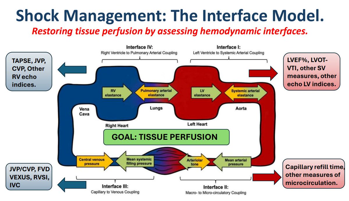
Image by peer reviewer Lewis McLean
Additional New Information
More on EMCrit
You Need an EMCrit Membership to see this content. Login here if you already have one.


