


[ad_1]
The following ankle x-rays are from a 65 year old female on steroids. She has fallen off a ladder and sustained an ankle injury. What can you see?

click to enlarge

click to enlarge

click to enlarge
[peekaboo_link name=”Answer”]Answer[/peekaboo_link]
[peekaboo_content name=”Answer”]
The ankle x-rays show diffuse osteopenia.
As shown below, there is a subtle calcaneal fracture. An important yet indirect sign leading to the diagnosis is a reduction of about 12 degrees or so in Bohler’s angle.

click to enlarge
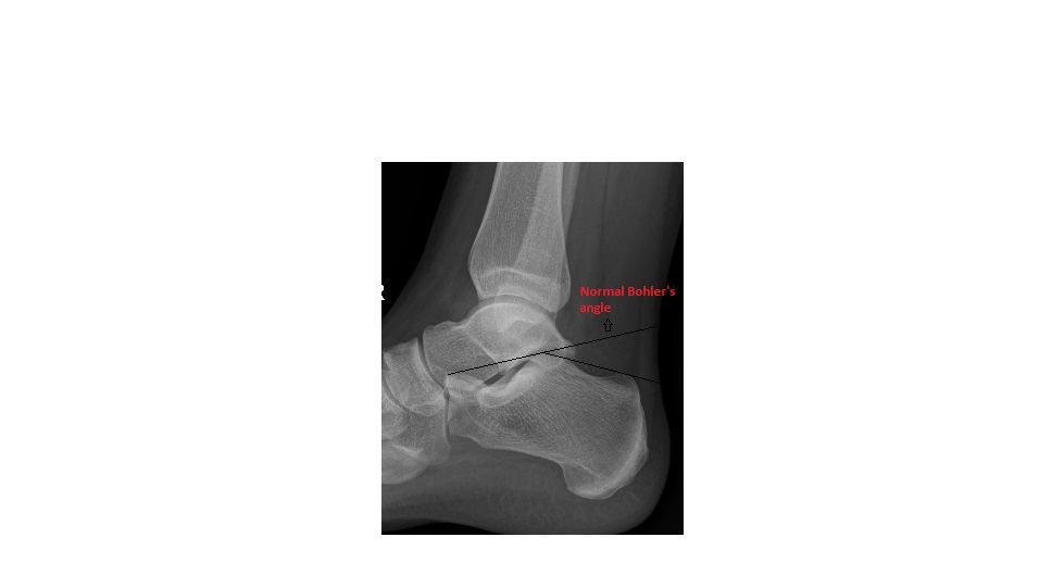
An ankle x-ray showing normal Bohler’s angle is shown below for comparison:
click to enlarge
Bohler’s angle is the angle formed between the:
- First line drawn from the highest posterior point of the calcaneum to the highest midpoint.
- Second line drawn from the highest midpoint of the calcaneum to the highest anterior point.
The normal angle should be 30-40 degrees. In calcaneal fractures, this angle gets reduced below 30 degrees due to the collapse of the cancellous bone in the calcaneum.
Sometimes, calcaneal fractures are only apparent when Bohler’s angle is measured.
[/peekaboo_content]
[ad_2]
Source link


