


[ad_1]
A 5 year old presents with FOOSH injury and a swollen elbow. His elbow x-rays are as follows. What do you notice?

click to enlarge

click to enlarge
[peekaboo_link name=”Answer”]Answer[/peekaboo_link]
[peekaboo_content name=”Answer”]
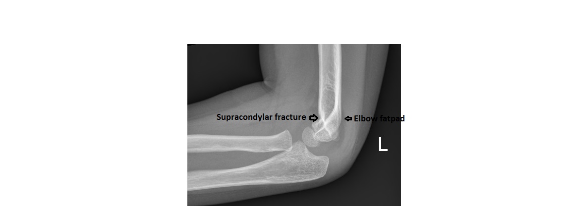
The AP view shows no obvious fracture and the radiocapitellar line is intact.
However, the lateral view shows the ‘sail sign’; the anterior humeral line has the entire capitulum lying in front of it. This should raise the possibility of a supracondylar fracture. If you look carefully, there is a supracondylar fracture with volar angulation of the distal humerus.
click to enlarge
More details on fat pad sign and anterior humeral lines can be found at:
http://www.emergucate.com/2012/10/08/imaging-case-of-the-week-16/
[/peekaboo_content]
[ad_2]
Source link


