


This 30 year old punched an object and heard a ‘click’ sound in his wrist following the episode. The following x-rays belong to the patient; what can you see?

click to enlarge

click to enlarge
[peekaboo_link name=”Answer”]Answer[/peekaboo_link]
[peekaboo_content name=”Answer”]
The xrays show no fracture around the wrist. The main abnormality is the ‘Terry Thomas Sign’ with widened scapholunate interval to 4.5 mm (normal scapholunate distance should be about 2-3 mm), which is indicative of scapholunate dissociation.
The Terry Thomas sign is usually accompanied by a signet ring appearance of the scaphoid due to its foreshortening. But radial deviation (as above) or flexion of the wrist can also give the scaphoid a signet ring appearance.
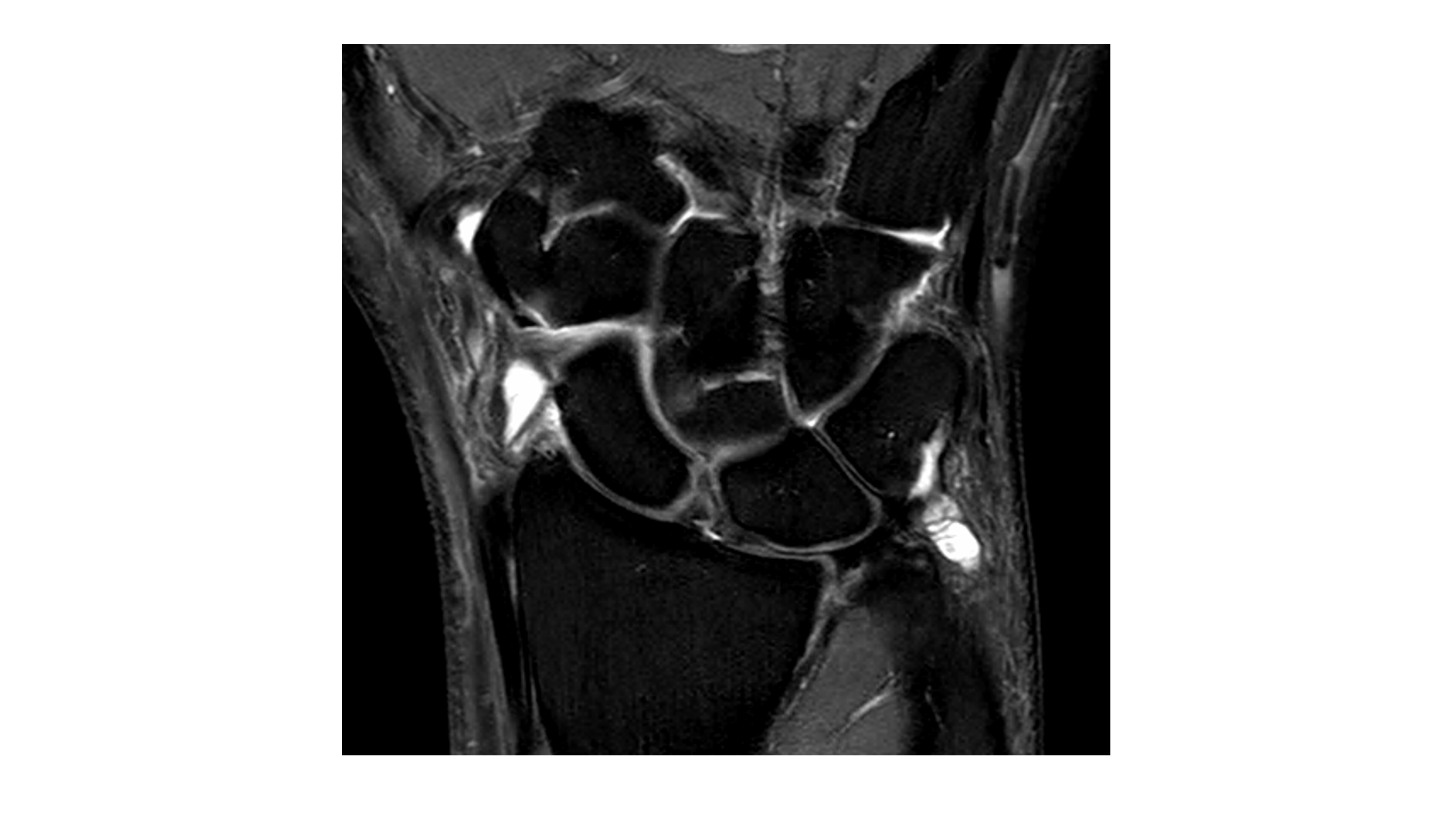
The following MRI from the same patient is a T2 fat suppressed sequence showing widening of the scapholunate gap.
click to enlarge
Scapholunate dissociation is an injury that can be commonly misinterpreted as a sprained wrist. This injury needs splinting of the wrist and a referral to hand surgeons.
[/peekaboo_content]


