


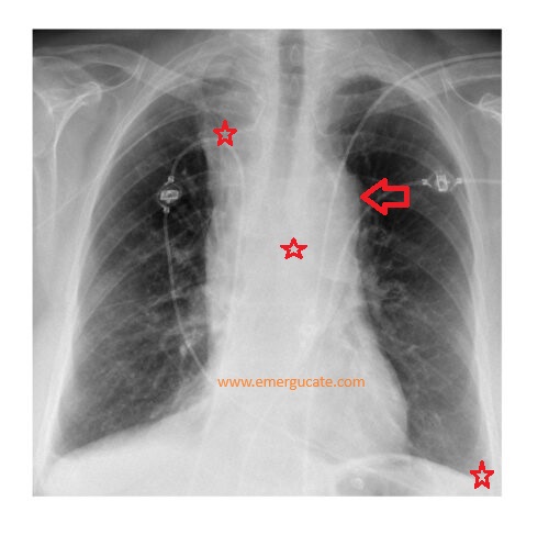
The chest x-ray shows features highly suggestive of thoracic aortic dissection.
- Mediastinal widening.
- Indistinct aortic knuckle.
- Right paratracheal density.
- Depression of the left main bronchus.
- Left pleural effusion.
Diagnosis was confirmed on CT aortogram and patient was referred urgently for specialist management.


